OCT & Optomap
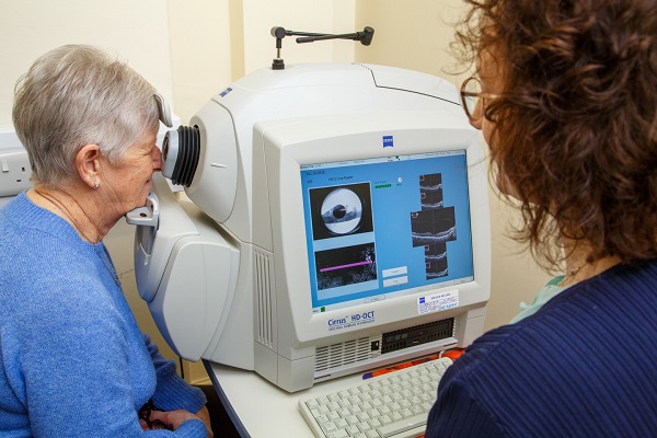
OCT (Optical Coherence Tomography) is a newish method of looking at the internal structure of the eye in great detail.
The technique is used both in hospital and optometric practice to view the retina, most often the macula, for signs of macular degeneration. In the case of the “wet” macular degeneration reveals the presence of fluid, and amount, in the retina This enables early diagnosis and treatment, and is used in the hospital setting to determine the length of treatment but also its efficacy.
If a person has glaucoma, then OCT is used to examine the structure that is most affected, the Optic Nerve Head. An OCT possess the ability to measure the thickness of the retinal nerve fibre layers around the nerve head and compare them to age related normal and also previous readings (where available) to see if there are any changes and thus monitor progression, or help in the diagnosis by showing if the fibre layer is thinner than normal.
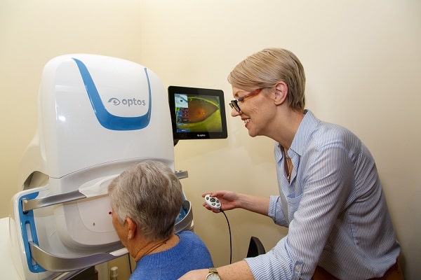
Optomap
The Optos Optomap is a form of camera which uses two different coloured laser beams to produce a wide field interior view of the eye. By sophisticated opto-electronics an Optos machine can produce a 200 degree retinal image. Most normal methods can only image a much smaller area, usually about 40 degrees per image (which may be able to be montaged with others to produce a larger image). In addition an Optomap can do this through a small undilated pupil, whereas the conventional camera often needs a dilated pupil (one that has been enlarged by the use of special eye drops) to do the same.
Corneal Topography
Corneal Topography is a special way of determining the shape of the cornea over its full extent. The normal method of measuring the curvature of the cornea known as Keratometry. Corneal Topography, however, measures the curvature over the whole cornea and presents a contour map, similar to an Ordnance Survey Map, of the corneal shape. This mapping is useful in diagnosing problems with the cornea, most notably Keratoconus. The advantage of this mapping is also demonstrated when special types of contact lenses are required to achieve optimal vision as the map can be used to generate a fully bespoke lens shape.